Cerebrospinal Fluid
Definition
Cerebrospinal fluid (CSF) is a clear, colorless body fluid found in the brain and spinal cord.
It is produced in the choroid plexuses of the ventricles of the brain.
There is also a connection from the subarachnoid space to the bony labyrinth of the inner ear via the perilymphatic duct where the perilymph is continuous with the cerebrospinal fluid.
Location
The CSF occupies the subarachnoid space (between the arachnoid mater and the pia mater) and the ventricular system around and inside the brain and spinal cord.
It constitutes the content of the ventricles, cisterns, and sulci of the brain, as well as the central canal of the spinal cord.
Contents
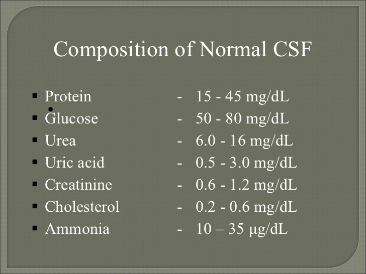
The CSF is derived from blood plasma
CSF contains approximately plasma proteins, approximately 15 to 40 mg/dL, depending on sampling site.
In general, globular proteins and albumin are in lower concentration in ventricular CSF compared to lumbar or cisternal fluid.
CSF is normally free of red blood cells, and at most contains only a few white blood cells.
Functions
It acts as a cushion or buffer for the brain
Provides basic mechanical and immunological protection to the brain inside the skull.
The CSF also serves a vital function in cerebral autoregulation of cerebral blood flow.
Buoyancy:
The actual mass of the human brain is about 14001500 grams
However, the net weight of the brain suspended in the CSF is equivalent to a mass of 25-50 grams.
The brain therefore exists in neutral buoyancy, which allows the brain to maintain its density without being impaired by its own weight, which would cut off blood supply and kill neurons in the lower sections without CSF.
Protection: CSF protects the brain tissue from injury when jolted or hit, by providing a fluid buffer that acts as a shock absorber from some forms of mechanical injury.
Prevention of brain ischemia:
Buoyancy facilitates blood perfusion.
Homeostasis:
CSF allows for regulation of the distribution of substances between cells of the brain, and neuroendocrine factors, to which slight changes can cause problems or damage to the nervous system. For example, high glycine concentration disrupts temperature and blood pressure control, and high CSF pH causes dizziness and syncope.
Clearing waste:
CSF allows for the removal of waste products from the brain, and is critical in the brain's lymphatic system.
Metabolic waste products diffuse rapidly into the CSF and are removed into the bloodstream as CSF is absorbed.
Production
The brain produces roughly 500 mL of cerebrospinal fluid per day.
It is constantly reabsorbed, so that only 125150 mL is present at any one time.
Produced by the choroid plexus, ependymal cells which line the ventricles and the lining surrounding the subarachnoid space.
Choroid plexuses also secrete growth factors, vitamins B1, 12 C, folate, beta-2 microglobulin, arginine vasopressin and nitrous oxide into the CSF.
A Na-K-Cl cotransporter and Na/K ATPase found on the surface of the choroid endothelium, appears to play a role in regulating CSF secretion and composition.
Reabsorption
CSF returns to the vascular system by entering the dural venous sinuses via arachnoid granulations.
There are valves to ensure one-way drainage.
Also reabsorbed through the sheathes of cranial and spinal nerves and through the ependyma.
Circulation
CSF circulates within the ventricular system of the brain.
From the ventricles the CSF passes through the interventricular foramina to the third ventricle
Then the cerebral aqueduct to the fourth ventricle.
From the fourth ventricle, the fluid passes into the subarachnoid space through four openings the central canal of the spinal cord, the median aperture, and the two lateral apertures.
There is a connection from the subarachnoid space to the bony labyrinth of the inner ear making the cerebrospinal fluid continuous with the perilymph.
CSF moves in a single outward direction from the ventricles, but multidirectionally in the subarachnoid space.
Fluid movement is pulsatile,
Abnormalities / clinical significance
Blood: Blood may be spilled into the CSF by accidental puncture of a leptomeningeal vein during entry of the LP needle.
Subarachnoid hemorrhage.
A few neutrophils and mononuclear cells may also be present as a result of meningeal irritation.
Xanthochromia (blonde color) of the CSF following subarachnoid hemorrhage is due to oxyhemoglobin which appears in 4 to 6 hours and bilirubin which appears in two days. Xanthochromia may also be seen with hemorrhagic infarcts, brain tumors, and jaundice.
Increased inflammatory cells (pleocytosis) :
Polymorphonuclear pleocytosis indicates acute suppurative meningitis.
Mononuclear cells are seen in viral infections (meningoencephalitis, aseptic meningitis), syphilis, neuroborreliosis, tuberculous meningitis, multiple sclerosis, brain abscess and brain tumors.
Leukemic cells in the CSF
Tumor cells indicate dissemination of metastatic or primary brain tumors in the subarachnoid space. The most common among the latter is medulloblastoma.
Increased protein: In bacterial meningitis, CSF protein may rise to 500 mg/dl.
Severe increase occurs in the Guillain-Barré syndrome.
Low glucose in CSF is seen in suppurative, tuberculous and fungal infections, sarcoidosis, and meningeal dissemination of tumors.
* * * * * * * * * * *